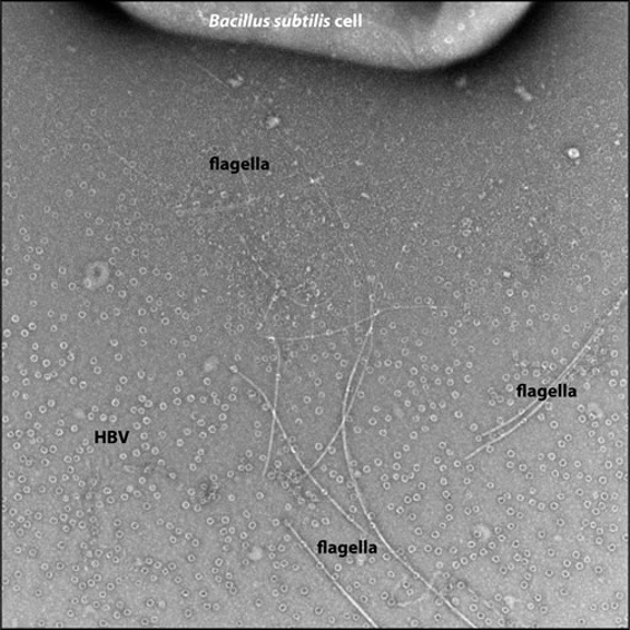
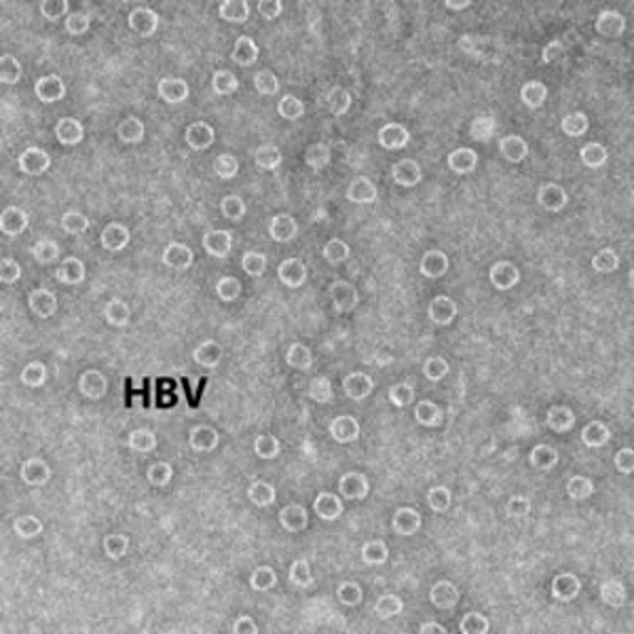
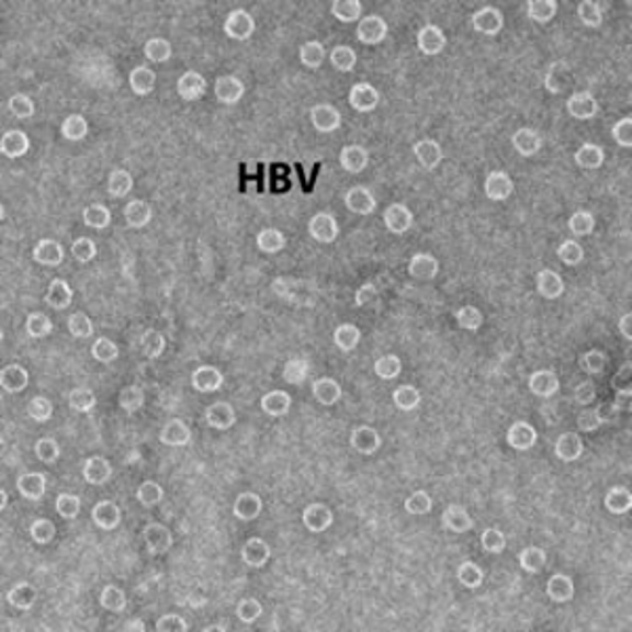
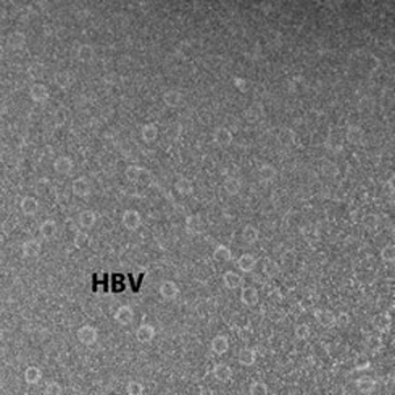
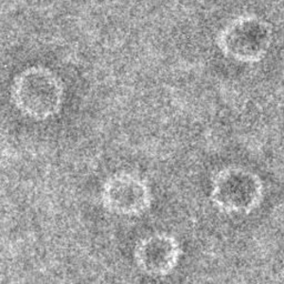
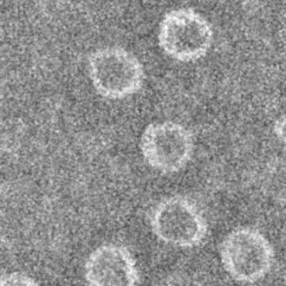
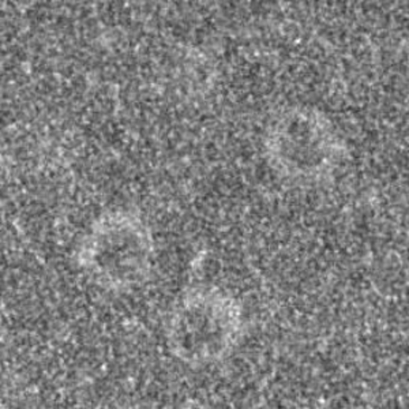
Some people (including reviewers of your papers!) are astonished by how different images of the same isolated material, stained under exactly the same conditions can appear. However, this is a quite common occurrence, has many causes and can even be seen in a single image (as illustrated using the image to the right, recorded using our JEOL JEM 3200FS). The image shows an edge of a Bacillus subtilis bacterium (along the top) with attached flagella that extend to the bottom edge of the image. The cell sits on a carbon film covered with hepatitis B virus capsids (HBV) and everything was stained with 1% uranyl acetate. Even in this single image, the appearance of the HBV and flagella changes dramatically from the bottom of the image to the top, as illustrated in more detail in the panel shown below of close-up images from three different regions of the image.
The left column shows a pair of close-ups of the HBV capsids from the lower left corner of the first image, where the lower image is a blow-up of the region around the HBV label in the top image. The middle column shows the same sort of image pair from the region of the original image that surrounds its "HBV" label. There is a fairly subtle difference in these two columns of images, where the contrast against the carbon support film is lower in the left column (and there is perhaps a bit more detail seen in the capsids in images from the middle column). The images in the right column (from much closer to the bacterium) are strikingly different: the overall contrast is much lower (i.e., the HBV capsids are much harder to see against the carbon support film and there is a nearly invisible flagellum running from upper left to lower right across the top image in this column) and little detail is visible in the individual HBV capsids.


