Defocus-Induced Magnification Changes

Defocus-Induced Magnification Changes

Unless the electron beam is carefully adjusted to be absolutely parallel to the optical axis as it passes through the specimen (i.e., neither convergent nor divergent), defocus changes in an electron microscope can cause a change in the magnification of images recorded on either film or a CCD camera. This change can be large or small, depending upon how non-parallel the electron beam is, and is highly dependent on the actual instrument used. For example, the JEOL JEM 1010 does not seem to exhibit this effect while the JEOL JEM 3200FS shows it readily.

The following graph illustrates this effect at a magnification of 12,000x using the JEOL JEM 3200FS:

A graph illustrating the effect of magnification at 12,000x which shows a linear trend.

This graph was generated from data obtained using serialEM and a standard replica diffraction grating to calibrate the pixel size, and no attempt was made to make the electron beam parallel. The graph clearly illustrates that a 1% change in magnification can occur when as little as 2 µm from focus, that this effect is equal whether the defocus difference is in the over-focus or the under-focus direction and that the effect seems to be linear, at least out to 10 or 15 µm from focus.

The following graphs illustrate this effect (again using the 3200FS) at different magnifications and different beam conditions (parallel or near-parallel, deliberately convergent, deliberately divergent, etc.).

NOTE: When indicating that the beam has been made "parallel" or "near-parallel," the following procedure has been used:

  1. switch the 3200FS into diffraction mode, then insert and roughly center any of the in-gap (OLA) apertures.
  2. focus the edge of the aperture to be razor sharp using diffraction focus.
  3. focus the unscattered beam in the diffraction pattern to as small a spot as possible using Brightness (CL 3).

This quick procedure sets the beam quite close to parallel. However, since the OLA does not sit exactly at the back focal plane of the objective lens, it will not ever make the beam exactly parallel and more complicated procedures should be followed.

Using 15,000x Magnification

Set beam close to parallel as it passes through the specimen, measure pixel size as a function of defocus using serialEM and a replica diffraction grating (waffle grid) and then make the beam more divergent and repeat the measurements.

This graph shows the dependent changes in pixel sizes at 15,000x magnification - the trend shows that the parallel beam fills the CCD and was unable to make the beam convergent.

The change in pixel size when the beam is close to parallel (red +'s) as it passes through the specimen is ~3% over a defocus change of ± 15 μm. This is about half the effect seen in the first graph, where no attempt to make the beam parallel was made. There is also some uncertainty (~1.9%) in the pixel size at the nominal defocus of "zero" that is likely due to both the serialEM procedure used and imperfections in the waffle grid. However, the trends are what is most important here: there is a small but very linear change in measured pixel size as the defocus is changed from -15 μm to +15 μm.

When the beam is made more divergent (i.e., when the diameter of the beam on the large focusing screen is expanded relative to the parallel condition, marked with green x's and blue ⁕'s in the above graph), this change in pixel size increases to more than 10% over that same ± 15 μm defocus range (and again, this change is linear and fairly smooth). The green x's and blue ⁕'s represent different amounts of divergence in the electron beam, with the blue ⁕'s showing data obtained with a more divergent beam. A line fit to the blue ⁕'s would be slightly steeper than one fit to the green x's, but it is not clear from this single pair of lines how the slope behaves as a function of beam divergence. Suffice it to say that when the beam is divergent, significant changes in pixel size can occur.

A more convergent beam should show similar changes in pixel size (but in the opposite direction - see below) but at 15,000x, the parallel beam just fills the CCD and it was not possible to make the beam convergent and repeat these measurements.

Using 50,000x Magnification

Set beam close to parallel as it passes through the specimen, measure pixel size as a function of defocus using serialEM and a waffle grid and then make the beam more divergent or convergent and repeat the pixel size measurements. At this magnification, it was necessary to use serialEM's montaging capability to image enough of the waffle grid to obtain an accurate measurement of pixel size.

This graph shows the dependent changes in pixel sizes at 50,000x magnification - the trend shows that the quality of the mantages will be effected by the defocusing effects.

The variation in the data here is much larger than at the lower magnifications, perhaps reflecting the fact that it was necessary to montage images in order to obtain enough repeats of the waffle grid to allow serialEM to determine the pixel size. It is also important to consider that serialEM's montaging process depends at least to some extent on the pixel size, and that as the defocus affects the pixel size, the quality of the montages will also be effected. Indeed, at many of the larger defocus values shown in this graph, serialEM had problems doing a montage, and those problems, in turn, prevented serialEM from determining the pixel size in at least some cases.

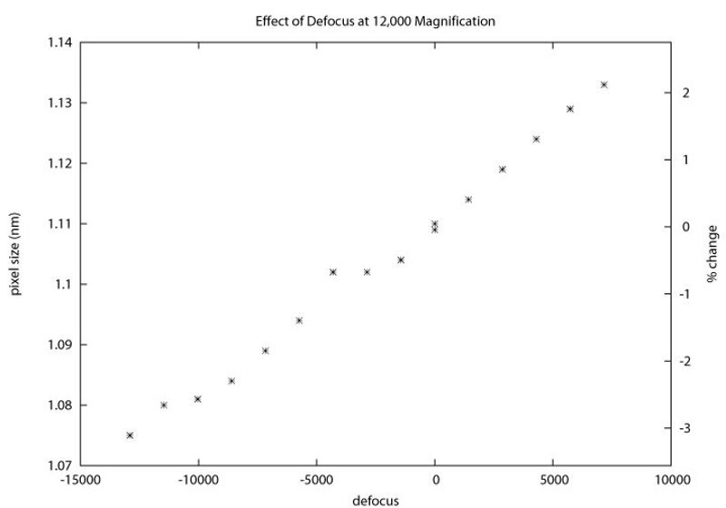
However, even with these data as messy as they are, the overall trends shown at lower magnifications remain true: when the electron beam is near-parallel (red +'s), there are relatively small changes in the pixel size (on the order of 3%) as the defocus is changed from -13 μm to +12 μm. When the beam is made divergent (blue ⁕), the change in pixel size grows to ~9% over a defocus range of -15 μm to +14 μm. As was also seen at the lower magnifications, when the beam is divergent, the pixel size increases as the images become more and more over-focused. When the beam is made convergent (green x's), the change in pixel size is ~8% over a defocus range of -10 μm to +7.5 μm. However, when the beam is convergent, the pixel size decreases as the images become more and more over-focused.