Cryo Electron Tomography

Cryo Electron Tomography (cryoET)

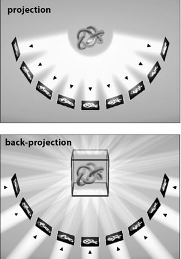
Electron tomography (ET) is a method that allows the user to generate a three-dimensional (3d) model of a specimen in the electron microscope by recording a series of images as the specimen is tilted along an axis normal to the electron beam. Most transmission electron microscopy (TEM) images are, at least to a first approximation, two-dimensional (2d) projections of the 3d specimen and several completely different formalisms have been used to show that it is possible to reconstruct a 3d object from 2d projections. For example, the figure to the left shows the relationship between projection images of a 3d object (top panel) and the back-projection of those 2d images to create a 3d volume (bottom panel). The different formalisms are the mathematical basis for the field of ET, and in recent years, the introduction of computer control over electron microscopes has made the technique available to most of the electron microscopy community.

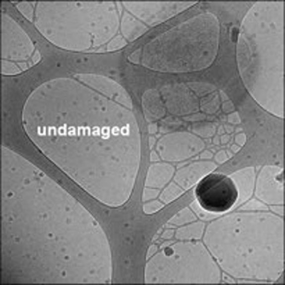
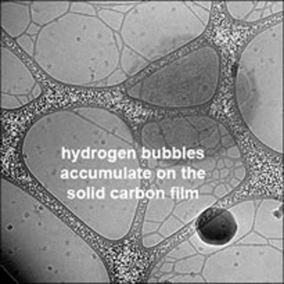
By severely limiting the electron dose, ET can be performed with a frozen specimen. This approach is called cryo electron tomography (cryoET) and simply merges extremely minimal dose ET data collection techniques with the use of a frozen sample. The sample can be a conventionally plunge frozen grid of the sort used for single particle imaging and reconstruction (and this is the type of sample most commonly used for sub-tomogram averaging), it can be a cryo-section cut from a much larger block of frozen material (e.g., a high pressure frozen sample) using a cryo ultra-microtome or even a cryo-section cut out of larger frozen block using a focused ion beam that operates at low temperatures (aka, a cryo-FIB). For the purposes of this discussion, specimen origins and properties will be ignored except in cases where it makes an important difference.

Reducing the Dose

The rule-of-thumb in much of cryoTEM work is that the total dose that the specimen receives should be limited to between 10 and 20 electrons/Å2. This is the level of exposure at which it is easily possible to measure high resolution structural damage in (for example) frozen, hydrated protein crystals inside a TEM. Isolated macromolecular complexes may (or may not) be somewhat more robust, but the very obvious bubbling, distortion and destruction of a frozen sample (e.g., figure on right) at slightly higher doses (50 to 100 electrons/Å2) indicates that damage is occurring at rates more or less comparable to what has been measured using frozen protein crystals.

It was thought for many years that the radiation sensitivity of such samples would preclude things like tomography, where in a typical tilt series, more than 100 images might be collected. Images at doses as low as (say) 10 electrons/Å2 are already so low contrast that identifying single macromolecular complexes can be hard in close-to-focus images, and recording more than 100 such images would produce a massive total dose (on the order of 1000 electrons/Å2). On the other hand, lowering the dose per image to ~1 electron/Å2 does lower the total dose to ~100 electrons/Å2 (which might be tolerable for low resolution reconstructions) but it was thought that the individual images would be so statistically ill-defined as to be both un-alignable (necessary for a 3d reconstruction) and generally useless.

However, more than 50 years ago, Reiner Hegerl and Walter Hoppe introduced a critical theorem stating that "A three-dimensional reconstruction requires the same integral dose as a conventional two-dimensional micrograph provided that the level of significance and the resolution are identical".

NOTE: The quotation comes from McEwen et al. (1995) Ultramicroscopy 60, 357-373 which cites Hegerl and Hoppe (1976) Z. Naturforsch. 31a, 1717-1721.

Another description of the idea behind this work is "dose fractionation" (i.e., the distribution of the electron (or X-ray) dose required to produce a statistically significant 3d reconstruction can be distributed over a (very) large number of individual images, each of which is itself statistically insignificant). In other words, provided that the total electron dose is sufficient to produce a statistically significant 3d reconstruction while also being low enough to avoid massive radiation damage, individual images can be extremely low dose (low signal-to-nose, statistically insignificant, etc.) provided that there is enough signal present to align the images to each other. Bruce McEwen, Ken Downing and Bob Glaeser published a paper in 1995 exploring the idea of dose fractionation using numerical simulations (the quotation above comes from the paper), and shortly after its publication, various groups around the world began the work that became the field now called cryoET.

As mentioned above, the key to cryoET is to limit the total electron dose to around 100 electrons/Å2. Doses this low mean that the dose per image should be 1-2 electrons/Å2 (depending on the number of images in a given tilt series). This should be the starting taget dose, but in practice, it may be necessary to decrease the total dose (either by recording fewer images or by lowering the dose per image). On the other hand, it may be possible to increase the total dose (by recording more images or by increasing the dose per image). The tolerable dose for any given specimen can range over a factor of 2 or 3, and...to be continued