There are times when the user of an electron microscope wants to make the electron beam either more or less intense as it hits the specimen. Additionally, the user may want to adjust the intensity by either a small or a significant amount. This is especially true when working with a frozen, hydrated biological sample that damages appreciably in the electron beam (i.e., where weak electron beams are required in order to limit the radiation damage), but is also useful in other situations. For example, when recording electron diffraction patterns, the beam should be bright enough to see the diffracted electrons well, but dim enough that the undiffracted electrons do not overload the recording medium (either film or some sort of CCD). It is also very important to keep in mind that while unstained biological samples damage quickly in the electron beam, virtually every sample will eventually damage if left in the electron beam long enough.
View some striking examples of beam damage
The most obvious way to adjust the intensity of the electron beam as it interacts with the specimen is to increase or decrease the area of the specimen that is illuminated. Since the beam current (the number of electrons passing through the condenser aperture per unit time) is constant as the size of the electron beam on the specimen is changed, increases or decreases in the diameter of the beam have a very strong effect on the number of electrons per unit area that hit the specimen. As an example, increasing the diameter of the beam by a factor of two increases the area of the specimen that the beam illuminates by a factor of four, and reduces the number of electrons per unit area by that same factor of four. However, there are circumstances when simply adjusting the diameter of the electron beam is not useful.
Several additional ways to alter the intensity of the electron beam before it interacts with the specimen are illustrated below using the Electron Microscopy Center's JEOL JEM 3200FS to adjust and measure the beam intensity and the Gatan Ultrascan 4000 CCD camera to record images under the different conditions. Different TEM's will behave slightly differently, but the effects illustrated here should also occur on all other instruments.
When recording images, the easiest way to control the electron dose in a single image is simply to vary the exposure time. This commonly happens when a user is trying to set a particular dose (in terms of electrons per Å2, for example) or when the beam is too bright or too dim for a camera to produce a useful image. Our data (exposure time vs the average value in a blank CCD image recorded with the beam covering the large phosphor screen at 25,000x) shows the effect of halving the exposure time, starting at 1 s and ending at 0.0625 s (a total of four steps). This is not an absolutely linear effect (the actual image counts are slightly less than expected based simply on reducing the exposure time by factors of two), but for the Gatan UltraScan4000 camera it is nevertheless quite good. Also be aware that longer exposure times result in a similar quasi-linear extension of the data shown in this plot. However, images recorded using long(er) exposure times can be degraded by relatively small amounts of specimen drift or vibration, especially at the high magnifications used for materials science work. It is also important to realize that adjusting the exposure time affects only the recorded image, and does nothing to influence the total electron dose the specimen experiences.
NOTE: Although the UltraScan 4000 on the 3200FS and the MegaScan795 on the JEOL JEM 1010 behave in the fashion described above, this is not true for all digital cameras. For example, the Gatan OneView on the EMC's JEOL JEM 1400plus shows rather different behavior.
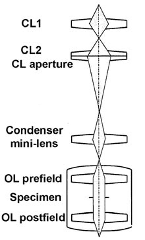
Another way to explicitly adjust the electron beam is to change the spot size setting on the microscope. Spot size settings control the 1st and 2nd condenser lenses (see diagram at top of page), and changes in spot size (from smaller numbers to larger numbers) result in the beam growing less and less intense (i.e., fewer and fewer electrons are passing through the condenser aperature above the specimen). Our data shows the effect (measured as the current density with the beam filling the large phosphor screen on our 3200FS at 25,000x) as a function of spot size.
The change is not linear, and is not even consistent from spot size to spot size (i.e., the amount of change in beam intensity from spot size 1 to spot size 2 is different from the amount of change from spot size 4 to spot size 5). However, a useful rule of thumb is that each sequential change in spot size is approximately a 2-fold change. If this were exactly true across all the different spot sizes, one would expect that the change from spot size 1 to spot size 5 would be a reduction to about 6.25% of the intensity of spot size 1 (a reduction to 1/24 of the intensity at spot size 1). In reality, this change is a reduction to ~10% of the original intensity.
Another way to influence the brightness of the electron beam is to change the condenser aperature (CL aperture in the diagram at the top of the page, corresponding to the CLA controls on our JEOL 3200FS): as the diameter of the condenser aperture grows smaller, the number of electrons passed through to the specimen also grows smaller (resulting in a less intense electron beam and a dimmer image). Our data shows the current density (recorded with the beam covering the large phosphor screen at 50,000x) as a function of condenser aperture position (where the largest aperture is at position 1 and the smallest at position 4).
Note: as of the end of 2014, the apertures in our 3200FS have diameters of 150, 70, 50 and 20 μm (CLA positions 1 thru 4).
Also note that in order to obtain the data for this plot, the magnification had to be raised from 25,000x (used for the other plots shown on this page) to 50,000x. The was needed because it was not possible to spread the electron beam to cover the large phosphor screen at 25,000x using the aperature in the 4th position.
It is also possible to combine changes in spot size with the different condenser apertures, resulting in a tremendous amount of control over the electron beam as it interacts with the specimen. The plot to the left shows counts per pixel in a 0.25 s exposure at 30,000x vs the beam current measured using a Faraday cup. The red +'s are condenser aperature 1, spot sizes 1 thru 5 (right to left, decreasing in both current and counts), the green x's are the same sort of data from condenser aperture 2, the blue asterisks are the same sort of data from condenser aperature 3 and the magenta boxes are the same data for condenser aperature 4. As the plot clearly shows, the beam current extends over more than 2.5 orders of magnitude and there are regions of overlap in the beam current where (for example) the beam with spot size 1, condenser aperture 3 can be brighter than spot size 5, condenser aperture 2. The counts per pixel shown in this plot were recorded with the beam spread to be completely visible within the CCD image using a 0.25 s exposure and a magnification of 30,000x. The high end of the data in this plot is at the point where increasing the number of counts/pixel much further will saturate (and can thus potentially damage) the sensor of the CCD camera.
With regard to the overlap between the beam current range of the sequential condenser aperatures, there may even be advantages to adjusting the electron beam to the same or a similar intensity using different combinations of spot size and aperture for different purposes. In addition, through careful use of what JEOL calls free lens control, it is possible for a user to adjust the beam intensity by the creation and use spot sizes that fall between or outside the 1 thru 5 settings that are normally available on the 3200FS.
Finally, the adjustments described here are useful for different sorts of TEM imaging and for diffraction. There are also ways to adjust the intensity of the electron beam.


