For the most part, the materials science electron microscopy community divides the world into "biological/soft" and "non-biological/hard" categories, where the designation of hard means that the materials are resistant to radiation damage. This is a misnomer since everything will eventually damage in the electron beam, but the basic idea has served the mat sci community well for many years. One group of inorganic materials known to damage readily are the zeolites, where certain of them are quite fragile in the electron beam.
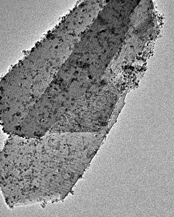
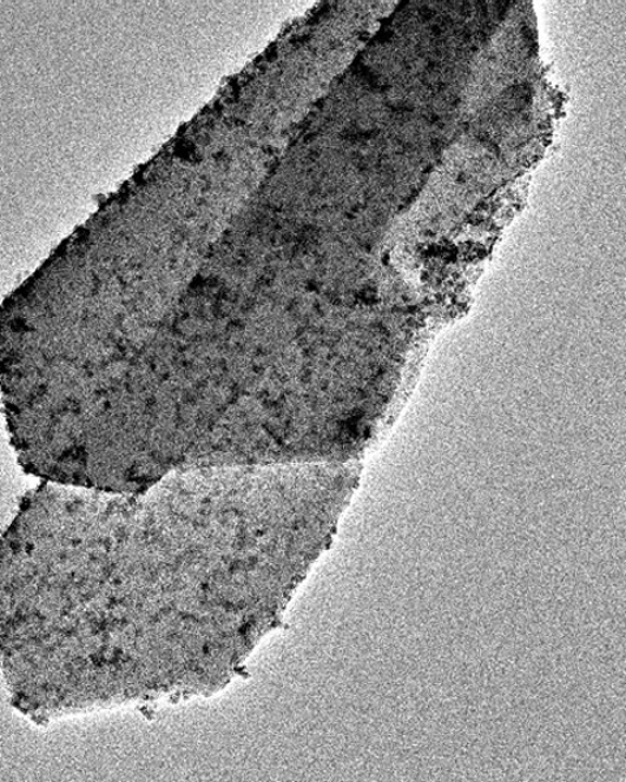
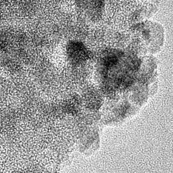
Here are a set of images demonstrating this fact. The image to the left was recorded early during a session on the JEOL JEM 3200FS and clearly shows large striations over most of the material that is indicative of the atomic order of this zeolite sample from Lyudmilla Bronstein's lab. The image to the right was recorded ~55 minutes later, after a series of very high magnification images of the Fe₃O₄ nanoparticles (dark particulates) that decorate this zeolite had been recorded. Even though the higher magnification images had been recorded at 8x higher magnification (400kx vs 50kx) with the illumination tightly focused onto much smaller regions of the zeolite, the atomic ordering (striations) seen in the first image have totally vanished from all the zeolite. Many details in the images have also changed due to exposure to the electron beam and resulting collapse of the zeolite.


