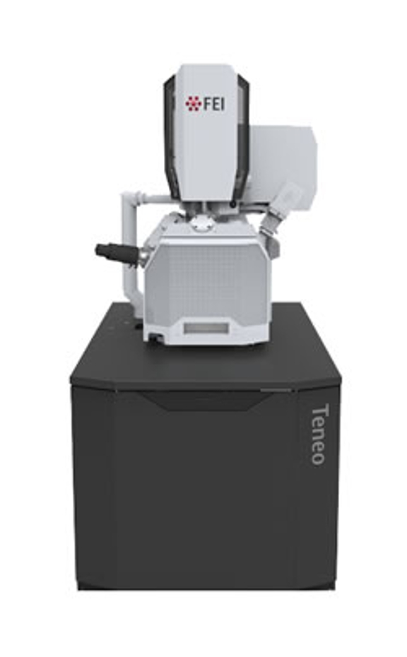
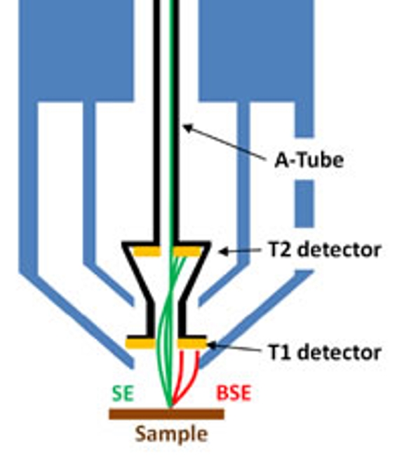
The Thermo-Fisher Teneo scanning electron microscope (SEM) was installed in Simon Hall 034 during January of 2018. The Teneo uses a field emission gun (FEG) electron source and can be operated at both high and low vacuum, using water vapor to adjust the chamber pressure from 10 to 50 Pa. It has an Everhart-Thornley detector (ETD), a low vacuum detector and two inside-the-pole-piece detectors referred to as T1 and T2. The T1 and T2 detectors are used to detect backscattered and secondary electrons, respectively.
The EMC also purchased the Thermo-Fisher VolumeScope attachment and software plugin to MAPS at the time the Teneo was purchased. The VolumeScope is essentially an ultramicrotome that functions inside the Teneo: as the ultramicrotome removes sections from a resin block, the Teneo scans the newly revealed blockface using the T1 (backscattered electron) detector. The ultramicrotome can cut sections as thin as 30 nm (what the VolumeScope refers to as the z-dimension) and the T1 detector can sample the blockface using steps as small as 5 to 10 nm in x and y. In addition, the VolumeScope can record SEM images at multiple accelerating voltages and perform energy deconvolution in a way that in effect allows the user to optically section into the blockface in steps finer than the ultramicrotome can cut.


