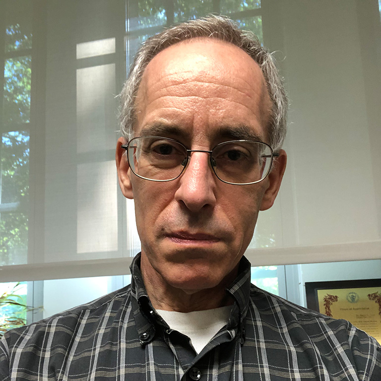
Barry Stein, Ph. D
Assistant Scientist
Myers Hall 040
812.855.7424
bstein@iu.edu
ORCID ID 0000-0003-2762-9434
I have 32 years of experience with electron microscopy, having started by using transmission electron microscopy (TEM) for my master’s degree research at Northeastern University and as a technician at the Massachusetts Institute of Technology, where I was in charge of the Center for Cancer Research electron microscopy laboratory.
At Northeastern University, I took the Biological Electron Microscopy course in 1981. The instructor for this was Dr. Daniel C. Scheirer. This class gave comprehensive training in the preparation of biological samples for TEM including fixation, dehydration, embedding, ultramicrotomy, staining, thin-film preparation, shadow casting, and carbon evaporation; along with the use and routine maintenance of an RCA EMU-3 TEM.
For my thesis research at NU, I worked in the laboratory of Dr. Michael Strauss between 1980 and 1984. The title of the thesis was “The leaf of taro, Colocasia esculenta (L.) Schott. Structure and ultrastructure”. The electron microscopes that I used were the Zeiss 9S TEM and the Amray 1000 SEM. I used both the LKB Ultramicrotome III and Sorvall Porter-Blum MT-2 ultramicrotomes to do the thin-sectioning.
When I was at the Massachusetts Institute of Technology, I worked for Dr. Phillip Sharp and was in charge of the electron microscopy laboratory at the Center for Cancer Research (CCR). Dr. Salvador Luria was the CCR director. I tutored graduate students in the use of a Philips 201 TEM, a Denton DV502 vacuum evaporator, and LKB Ultramicrotome III, as well as doing research. I also had access to equipment at the Whitehead Institute for Biomedical Research, where I used a Reichert Ultracut E with a FC-4 cryo unit attachment to prepare samples for immunolabeling. This work was done for Dr. Shiv Pahlavi in the laboratory of Dr. David Baltimore. I worked at MIT from 1983 to 1985.
I studied for my PhD at Michigan State University (MSU) between 1985 and 1991. While I was there, I was a teaching assistant for NSC-810, Transmission Electron Microscopy. This class was given by my dissertation adviser Dr. Karen Klomparens, who was director of the Center of Electron Optics at MSU, and by her research associate Dr. John Heckman. I was also a teaching assistant for NSC-820, Scanning Electron Microscopy, for Dr. Stan Flegler, who is now the director for the Center for Advanced Microscopy at MSU. Both of these classes were designed to give students the ability to conduct research using transmission or scanning electron microscopy. Each class would have four to ten students. My duties consisted of guiding the students on an individual basis through their assignment for that week. In addition, I helped those students who had already taken the course with their research. For TEM class, we would go through usage of the Phillips 210, photography, preparation of large and small samples in epoxy blocks for ultrathin sectioning, ultramicrotomy, preparation of coated grids, negative staining of samples, and metal shadow casting of particles mounted on coated grids. For the SEM, we went through usage of the JEOL 35CF, stigmation, the use of gamma in imaging, photography using a Polaroid camera, stereo photographs, preparation of large and small biological samples for SEM, sputter coating of the samples, working distance, adjustment of the spot size and accelerating voltage.
In my dissertation research, I examined the ultrastructural responses of cucumber plants to infection by the fungus Colletotrichum lagenarium. To do this, I used standard preparation techniques for TEM and SEM, including ultramicrotomy. I found that areas of apparent lignification, which has often been correlated with resistance in the literature, occurred in cell walls of the cucumber plants directly under areas of fungal infection.
To further examine lignification in cell walls, I stained the samples with potassium permanganate and bromine, both of which bind to lignin and cause an increase in the electron density of the lignin. This allowed me to localize lignin in the walls of cells that were adjacent to the sites of infection. To confirm that both bromine and manganese were present, I used a JEOL 100CX II TEM/SCAN with a Link Systems AN 10000 to prepare energy dispersive X-ray microanalysis (EDS) dot maps of the localization of the two elements.
In the same location as the areas of lignification, I also found that the cell walls contained silicon. In previous reports, plants that were given higher concentrations of silicon were reported to have increased resistance to infection but the use of EDS, showing the localization of silicon next to the pathogen, provided a reason for this effect.
This portion of my dissertation research was published in two papers: B.D. Stein, K.L. Klomparens, and R. Hammerschmidt (1992) Comparison of Bromine and Permanganate as Ultrastructural Stains for Lignin in Plants Infected by the Fungus Colletotrichum lagenarium Microscopy Research and Technique 23:201-206 and B.D. Stein, K.L. Klomparens, and R. Hammerschmidt (1993) Histochemistry and Ultrastructure of the Induced Resistance Response of Cucumber Plants to Colletotrichum lagenarium Journal of Phytopathology 137:177-188.
For my dissertation research I also used double jet propane freezing to prepare samples for use in a Balzers freeze-fracture/freeze etch unit.
While a postdoctoral researcher at Michigan State University and the Ohio State University I used immunogold labeling of plant and animal tissue using samples embedded in Lowicryl, London Resin (LR) White, and LR Gold. For these research projects, the transmission electron microscopes that I used were a Zeiss 10 and a Philips CM-10.
As a research associate with Dr. Saul Tzipori at the Tufts University School of Veterinary Medicine, I used both a Philips CM-10 and a JEOL 1010 TEM, to examine epoxy embedded samples prepared by standard techniques and immunogold labeled LR White embedded samples, and an ISI DS130 scanning electron microscope.
I was the manager of the Indiana Molecular Biology Institute microscopy facility in Myers Hall 040 starting in July 2000. This facility and the Simon Hall EM facility were merged in July 2013 to become the current Electron Microscopy Center. I train and supervise users of the JEM 1010 TEM and JSM 5800LV SEM and instruct the users in sample preparation techniques along with the use of the equipment that is needed for this, including ultramicrotomes, vacuum evaporator, sputter coater, and critical point dryer. Once a year in the spring semester, I participate in teaching, with Sid Shaw, Jim Powers, and David Morgan, a Z620 course on light and electron microscopy. I participate in two other courses, Cell Biology Laboratory L313 and Viral Tissue Culture Laboratory M435 where, during one of the class sessions, I demonstrate the electron microscopes. My research collaborations have resulted in my being included as a co-author on 67 refereed journal articles. I have been a reviewer for Veterinary Microbiology, as well as several other journals, since 2008.


