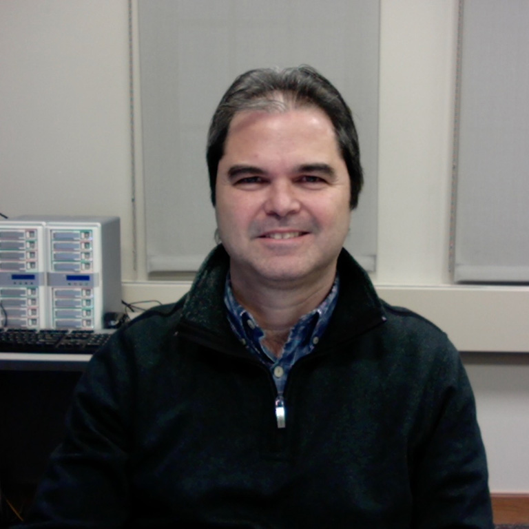
Giovanni Gonzalez-Gutierrez, Ph. D
Assistant Scientist
Simon Hal 005A
812.856.7505
giovgonz@iu.edu
ORCID ID 0000-0002-1044-943X
Throughout my scientific career I have approached several fundamental biological questions in living cells. During my early career years, I aimed to study the coupling of energy utilization to the conduction of ions through the cell membrane and how this phenomenon is mediated by ion channels and other regulators.
Furthermore, during my years at the Indiana University, I have used the power of X-ray crystallography and more recently single particle Cryo-EM, to elucidate the structural mechanisms of several enzymes as well as to unveil potential new therapeutic strategies in cancer research by using small molecules covalently bound to proteins involved in tumor proliferation.
The relevance of ion channels and transporters in many psychological and neuronal diseases is unarguable. One third of all drugs in the market actually target several members of these membrane expressed proteins. The superfamily of voltage-gated ion channels and the superfamily of neurotransmitter Cys-loop receptors, which I study in molecular detail, are represented by several members. For instance, the voltage-gated calcium channel is widely expressed in brain, specifically in the synaptic terminals of neurons. Calcium inward current triggers the release of neurotransmitters, a fundamental biological mechanism in interneuron communication. Additionally, calcium channels play a very relevant role in skeletal and cardiac muscle during contraction. Under the supervision of my doctoral advisors Dr. Alan Neely and Patricia Hidalgo, my research pinpointed the role of the beta-subunit of the voltage-dependent calcium channels as the most important regulator of the alpha1 pore-forming subunit. Despite the chaperone role played by the beta-subunit, I focused on the role of beta-subunit to modulate ion currents, voltage-dependence, and ability of the pore to open and close. I found that only the guanylate kinase like-domain of beta-subunit (and not the SH3 domain as previously suggested) is necessary to modulate all gating properties that are actually regulated by the full-length protein (Gonzalez-Gutierrez et al. PNAS 2010). This implies, that only the interaction of beta-subunit to the AID (with no additional secondary interactions) suffices to favor energetically the channel gating at lower voltage. Likewise, I demonstrated that the SH3 domain of beta-subunit plays a relevant role in the interaction with other intracellular proteins (i.e. dynamin) and in its turn, it can effectively down-regulate calcium channels from plasma membrane (Gonzalez-Gutierrez G et al. JBC 2007). Furthermore, I elegantly proved that the beta / alpha1 subunit interaction is reversible and the free full-length beta-subunit can also down-regulate calcium channels (Hidalgo et al. JBC 2006). Altogether, these studies enlighten the role of calcium channel regulators in cell physiology.
My work during the postdoctoral training, in the Department of Molecular Integrative Physiology at the University of Illinois at Urbana-Champaign, under the supervision of Dr. Claudio Grosman focused on the structure-function of Cys-loop receptors. These receptors are represented by several members, also expressed widely in the brain, specifically in post-synaptic membranes. Among Cys-loop receptors are the acetylcholine nicotinic receptor, the 5HT3 serotonin receptor, the GABA receptor and the glycine receptor. These receptors are linked to devastating neurological diseases such as: Alzheimer’s, epilepsy, schizophrenia, addiction and others. My structural studies on bacterial members of the superfamily demonstrated that caution must be followed while assigning functional states to structural models (Gonzalez-Gutierrez G et al. JMB 2010; Gonzalez-Gutierrez G et al. PNAS 2012; (Gonzalez-Gutierrez G et al. JGP 2017). I have also obtained structural models for several end-states of the gating pathway, for instance the fully-liganded close conformation and stabilized open-conformation in the same system (Gonzalez-Gutierrez et al. PNAS 2013; Gonzalez-Gutierrez G et al. JGP 2017). This is especially relevant to get insights about the mechanism of gating in these ion channels. Likewise, I have made the functional characterization of the bacterial members of the superfamily, which are more amenable to structural studies (Gonzalez-Gutierrez G et al. JMB 2010; Gonzalez-Gutierrez G et al. JGP 2015).
More recently at the Indiana University, I have been involved in several collaborative projects. This has been aside my duties as facility manager in both the Automated Crystallography Facility (CAF) and the Physical and Biochemistry Instrumentation Facility (PBIF), a complementary function that I really enjoy as it involves much more interaction with PhD students, postdocs and scientists on campus. In association with Dr. Samy Meroueh, I have solved the structure of human TEA domain (TEAD) transcription factors with a small molecule that rapidly and selectively forms a covalent bond with a cysteine in the palmitate binding pocket (Bum-Erdene K et al. Cell Chem. Biol. 2019). TEAD transcription factor is activated ultimately by binding with YAP after the Hippo signaling pathway has been triggered. It’s well-established that the pathway regulates cell proliferation and tumor progression. Therefore, these small molecules can be potentially used in cancer therapeutics.
Jointly with Dr, Meroueh, we have discovered a new well-defined binding site in RalA (Ras-like) GTPase. I have solved five high-resolution structures (1.1 - 1.5 Å) of small molecules covalently bonded to a tyrosine residue in the above-mentioned binding site (Bum-Erdene K et al. PNAS 2020). Traditionally, cysteines have been the favorite target given the high reactivity and the ability to form covalent bonds in a wide range of conditions. However, cysteine residues are not always present in proteins (for example, RalA) or are not exposed enough in order to be reached by chemical compounds. Thus, our finding about the suitability of tyrosine residues as a target to form covalent bonds will definitely redirect the focus to the search of new therapeutical strategies in cancer research. New small-molecules-RalA crystal structures are currently being pursued.
My collaboration with Dr. David Giedroc has been also very successful and we have published several papers. With Jifei Wang, a former PhD student, I solved the structure of RibBX a protein involved with Flavin metabolism in Acinetobacter baumannii (Wang J et al. Cell Chem. Biol. 2019). Furthermore, I have solved the structures of several mutants of the chromosomal zinc-regulated repressor CzrA (Capdevila DA et al. JACS 2018). Moreover, I have solved the structures of reduced and oxidized forms of SqrR, a sulfide-responsive transcriptional repressor. SqrR acts as a regulator of sulfide-dependent gene expression and it’s able to form a tetrasulfide bond between two cysteines highly conserved in the family (Fig. 5). Remarkably, the crystal structure of the SqrR tetrasulfide form is to our knowledge, the first member in the PDB containing such a covalent bond.
I have similarly solved crystals structures for different groups at IU including, Dr. Stephen Bell, Dr. Hengyao Niu, Dr. Carl Bauer, Dr. Adam Zlotnick, Dr. Richard DiMarchi, and Dr. Julia van Kessel. With Dr. Bell, and Dr. Bauer, crystal structures of two new proteins were solved using single-wavelength anomalous dispersion on very weak anomalous data. With Dr. Zlotnick’s group we published the structure of the dimeric capsid protein (Cp) from woodchuck hepatitis virus (WHV), a hepatitis B virus homologue (Zhao Z et. al. J. Virol. 2019). Jointly with Dr. Piotr Mroz from DiMarchi group we solved the structures of nine variants of human glucagon at resolution of 1 Å (PDB codes 6PHI-Q). Lastly, with Dr. van Kessel’s group we solved the structure of five variants of SmcR from Vibrio vulnificus, a transcriptional regulator of the quorum-sensing regulon (PDB codes 6WAE-I). Given our current involvement with multiple projects, I foresee an increasing number of new structures and relevant questions to be answered at regular pace in the forthcoming years
The advent of single particle Cryo-EM in recent years has brought another tool to structural biologists. I am interested in solving large macromolecular complexes and develop new validation tools for Cryo-EM structural models.
List of PDB depositions:
6WAE 6WAF 6WAG 6WAH 6WAI 6P0I 6P0J 6P0K 6P0L 6P0M 6P0N 6P0O 6O8K, 6O8L, 6O8M, 6O8N, 6O8O, 6ECS, 6MNZ, 6E5G, 6CDA, 6CDB, 5V6N, 5V6O, 4LMJ, 4LMK, 4LML, 3UQ4, 3UQ5, 3UQ7
Complete list of Published Work in MyBibliography:
https://www.ncbi.nlm.nih.gov/myncbi/16sPivSdArzQb/bibliography/public/


